
Panoramic X-ray and 3D scanner
Are they dangerous?
Dental panoramic scanners and 3D dental scanners have significantly improved the predictability of the results of dental interventions, especially regarding surgical ones. However, with their appearance, the question arose whether these technologies are completely harmless to human health or can help the emergence of tumors and the development of cancer. Therefore, we will try to understand the relationship between the benefits and risks of this type of research, while describing in detail the precautions that must be taken to avoid future health problems.
Why do you need panoramic X-rays or 3D scanner diagnostics?
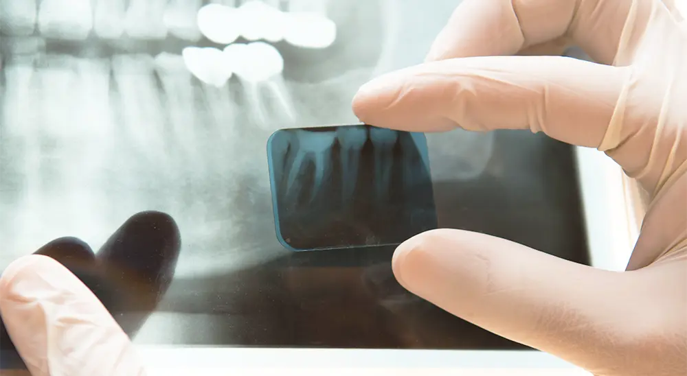
Panoramic X-ray (OPG) photography is an absolutely necessary study aimed at giving comprehensive information about the overall condition of the jaw, as it visualizes all the teeth, upper and lower jaw and paranasal (maxillary) sinuses. Thanks to the OPG photo, the doctor can understand the condition of all teeth, whether there are pathological changes in the bone, retained teeth, cysts, horizontal and vertical bone loss (periodontitis), temporomandibular joints (TMJ), mandibular canal, wisdom teeth and joints.
The 3D scanner is most often used as a method for successful planning in the placement of dental implants, as well as for 3D implantation. 3D scanner diagnostics is also used to determine the thickness and size of the bone, the exact location of the maxillary sinuses and their condition, the location of the mandibular canal for implantation or removal of wisdom teeth, the presence of granulomas, cysts, and periodontitis.
The 3D scanner allows you to model the entire jaw in three dimensions to simulate the intervention in a very precise way and to achieve the best possible results.
What are the risks of taking a panoramic X-ray and 3D scanner?
Every day each person receives a certain dose of radiation from the natural background. By taking a sector dental image, for example, the patient receives the dose of radiation that he would receive from the natural background within 1 day. This proves the claim that the ionizing radiation of X-rays used for dental purposes is very low.
It is estimated that taking a dental photo can be as dangerous as the chance of a car accident on a 100-kilometer journey.
The risk of radiation during an X-ray examination is smaller than the danger of not finding out the problem on time, which can lead to serious complications.
What are the precautions for performing dental X-rays?
In Europe, dental surgeons are required to attend regular radiology training courses to monitor the development of techniques and devices. Therefore, they are fully aware of the precautions that must be taken and do everything possible to protect the health of patients.
During the examination, for maximum patient safety, it is necessary to remove all metal objects from yourself (jewelry, accessories, etc.). The X-ray technician will then put on a lead vest and collar to protect you from radiation.
Can dental X-rays be performed on pregnant women?
Before proceeding with an X-ray examination, it is necessary for the patient to consult with her personal obstetrician-gynecologist and to inform the X-ray room in a timely manner. Dental X-rays can only be taken if it is necessary. The test is performed under a strict protocol – wearing a protective collar and a lead vest, which aims to protect both the mother and the fetus from radiation. With the measures taken in this way, the danger is minimized.