Why it is necessary to take an orthopantomography x-ray after endodontic treatment?
The orthopantomography scan for treatment with pulpitis and periodontitis or why is it necessary to make an orthopantomography, after the filling of tooth canals?
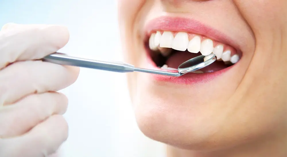
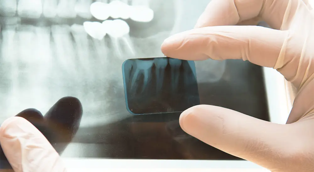
What is a dental X-ray scan?
Dental X-ray scan is a type of X-ray which examines the tooth and the bone around it, which helps the doctor grasp a full comprehensive idea regarding the whole clinical scan.
The most common use of dental x-ray scans is for Orthopantomography OPG (Panoramic x-ray picture) and Segment x-ray (segment radiography).
The information, which the doctor receives during a similar examination is definitely very important:
The orthopantomography is used for very complex diagnostics, which shows the status of all the teeth and all the pathological changes in the bone. The segmental x-ray gives much better information regarding the full status of the teeth and the bone, which surround them. They are mostly used when the patient has a specific complaint, connected to a specific tooth or with the control of an already completed procedure.
What is endodontic treatment and what is the protocol for its successful course?
The endodontic treatment (root treatment) is a procedure, aiming to eliminate the infection from or around the root canals. The treatment can end in one or a couple of visits, depending on the severity of the individual case.
First step:
It is extremely important to determine the length of the root canals, for the treatment to be successful. In most cases, this happens through an apex locator or segmental radiography. After the tooth is numbed, we place a dental dam, which isolates the tooth and protects it from saliva, during the whole process.
Second step:
Through an appropriate cavity, this provides access to the root canal which allows the root of the tooth to be used as an instrument (manual or mechanic) for removing the pulp and the root contents. After this, the roots are processed and given an appropriate shape.
Third step:
After the forming and cleaning, the canals are filled with a biocompatible material, most-commonly gutta parka and sealer.
Fourth step:
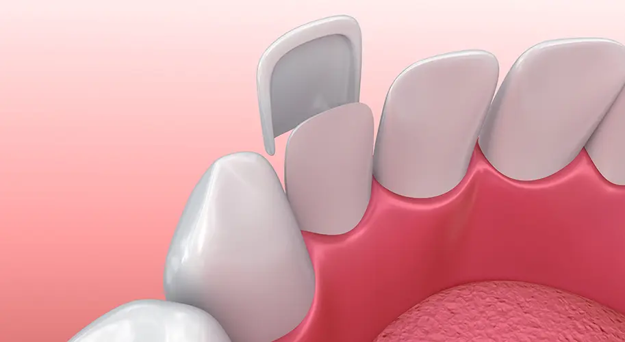
Before moving onto the restoration of the tooth with a crown or with obturation (filling), to provide the protection and restoration of its functions, it is recommended to make a control segment x-ray scan. This will give clear information to the dental doctor regarding whether the nerves have been fully removed and whether all the canals are filled.
The x-ray scan takes only one minute, the price is very small in comparison, to the information it provides.
In our practice very often, we encounter patients who have been treated for pulpits, who have not had any control x-ray scan pictures, and after filling the canals, it was necessary to treat the same tooth again because of that reason.
The absence of control x-ray scans very often leads to unnecessary consequences, which leads to spending more time with the dentist and spending more money on fillings (obturation).
Re-treatment of dead teeth requires the removal of any existing fillings (obturations), removal of the root stuffing and the renewal with new stuffing, this is to be respected throughout the whole protocol.
Re-treatment poses multiple risks and its success rate is under question. It is important to follow the protocol of treatment for pulpit which is confirmed with a control x-ray scan.
When the nerve has not to be removed completely, and the tooth is filled, this unremoved part necrotizes and can lead to gangrene and very often granuloma to pass into a cyst.
Very often all these diagnostics flow asymptomatically. Because of this reason, the patient finds out later about the problem and quite often this leads to the extraction of already ‘treated’ teeth, while all of this could have been avoided by taking a control orthopantomography scan.